Artigo: Proximal Humeral Intramedullary Nails: 5 Common Errors and How to Avoid Them
Proximal Humeral Intramedullary Nails: 5 Common Errors and How to Avoid Them
Proximal humeral fractures represent a constant challenge in orthopedic practice, especially in elderly patients with osteoporotic bone. Although locked plate fixation is widely used, the use of third-generation intramedullary nails (IMN) has gained prominence due to their biomechanical advantages and less invasive approach. However, the technique is demanding, and small errors can compromise the functional outcome.
Based on the guidelines of Rockwood & Green's Fractures in Adults (9th Edition), this article details the five most common errors in osteosynthesis with intramedullary nails in the proximal humerus and strategies to mitigate them.
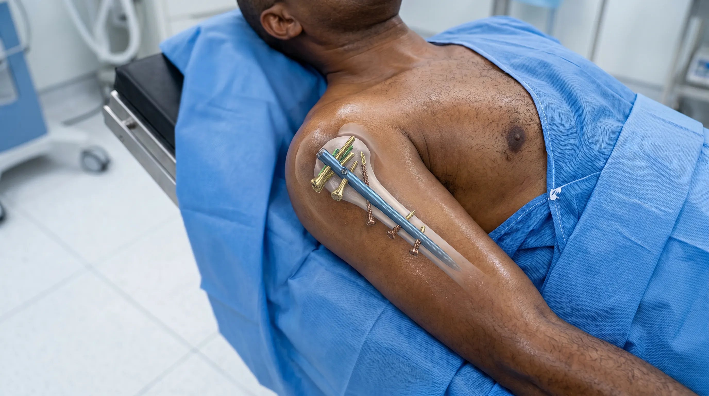
1 - Fracture Displacement During Nail Insertion
A common mistake is allowing nail insertion to displace a fracture that was already well reduced. This usually occurs when the entry point is not perfectly aligned with the medullary canal or when the temporary reduction is not stable enough.
- How to avoid: Reduction must be achieved and confirmed by fluoroscopy in two planes (AP and Y-profile) before preparing the entry site. The use of Kirschner wires or Schantz pins as "joysticks" outside the planned nail trajectory is essential to maintain alignment during reaming and insertion. The exact entry point depends on the nail design (curved vs. straight), and the use of contralateral templates can help predict the diameter and ideal point.
2 - Inadequate Entry Point and Rotator Cuff Injury
The choice of entry point is critical. Nails with lateral curvature (curvilinear) require a more lateral entry point, close to the rotator cuff insertion, which can increase the risk of chronic postoperative pain. Straight nails, on the other hand, require a more medial entry, through the musculotendinous junction or even the articular surface.
- How to avoid: To minimize damage, the incision in the supraspinatus tendon should be made cleanly and longitudinally to its fibers, preferably 1 to 1.5 cm posterior to the biceps tendon (a key anatomical landmark). The use of soft tissue protectors during reaming is mandatory to prevent rotator cuff "abrasion." In addition, careful closure of the cuff at the end of the procedure is essential for functional recovery.
3 - Prominent Nail Positioning (Impingement)
Leaving the proximal end of the nail too high is a serious technical error that leads to subacromial impingement, severely limiting abduction and causing pain.
- How to avoid: The nail must be countersunk at least 10 mm below the articular surface or the tip of the greater tuberosity. The depth must be rigorously checked under fluoroscopy before distal locking, ensuring no metallic prominence that could collide with the acromion during shoulder movement.
4 - Articular Penetration of Locking Screws
The spherical anatomy of the humeral head and poor bone quality facilitate perforation of the articular surface by proximal screws, either due to incorrect measurement or secondary fracture collapse (varus collapse).
- How to avoid: When measuring screw length, it is recommended to select a screw 5 mm shorter than measured when directed towards the articular surface. The use of screws with blunt tips and "screw-in-screw" technologies or polyethylene bushings (characteristics of 3rd generation nails) increases angular stability and reduces the risk of "back-out" or migration.
5 - Neglecting Tuberosity Fixation
In 3-part fractures, failure to adequately stabilize the tuberosities (especially the greater tuberosity) results in loss of external rotation and elevation strength. The nail alone may not provide sufficient rotational stability for these fragments.
- How to avoid: Use the proximal nail perforations that allow suture passage. Rockwood recommends using heavy traction sutures (such as #5 polyester or #2 polyethylene) passed through the tendon-bone junction. These sutures should be incorporated into the nail construct, creating a "tension band" that neutralizes the deforming forces of the rotator cuff.
Summary Table: Pitfalls and Preventive Measures (Proximal Humeral IMN)
| Common Error | Clinical Consequence | Prevention Strategy |
|---|---|---|
| Entry point too lateral | Rotator cuff injury / Chronic pain | Use biceps as reference; precise longitudinal incision. |
| Prominent nail | Subacromial impingement | Countersink nail >10mm below bone. |
| Long screws | Chondrolysis / Joint pain | Undersize screw by 5mm; blunt tips. |
| Insufficient reduction | Malunion / IMN failure | Anatomic reduction prior to reaming (joysticks). |
| Lack of tuberosity suturing | Rotator cuff dysfunction | Incorporate high-strength sutures into proximal locking. |
Conclusion
Osteosynthesis of proximal humeral fractures with intramedullary nails is a powerful technique, but it requires millimeter precision and a deep understanding of the technical pitfalls. By avoiding positioning errors, respecting the anatomy of the rotator cuff, and ensuring the stability of the tuberosities, the surgeon can offer the patient early rehabilitation and an excellent functional outcome.
The learning curve to master these nuances can be drastically reduced through observation of real surgeries with high technical quality and immersive perspective.
Master the Technique in Practice
To deepen your knowledge and visualize every detail of the application of these techniques in a real surgical environment, we invite you to watch the training courses on the Reconstrução Óssea platform. Check out the relevant content:
- Intramedullary Nail for Proximal Humerus (03): Master proximal humerus fixation with fluoroscopy-guided intramedullary nailing in 4K vision.
- Proximal Humeral Fracture in 2 Parts (Deltopectoral Approach): Training focused on reduction and fixation, essential for understanding anatomy and surgical alternatives.
- Reconstruction of Complex Four-Part Proximal Humeral Fracture: Advanced management of complex fractures with a focus on reconstruction and stability.
- Proximal Humeral Nail Technique (Blog Post): Detailed article accompanying the narrated surgery video, focusing on precision and safety.
Watch now and elevate the level of your surgical practice: https://reconstrucaoossea.com/
Reference: Court-Brown, C. M., et al. (2020). Rockwood and Green's Fractures in Adults. 9th Edition. Wolters Kluwer.
