
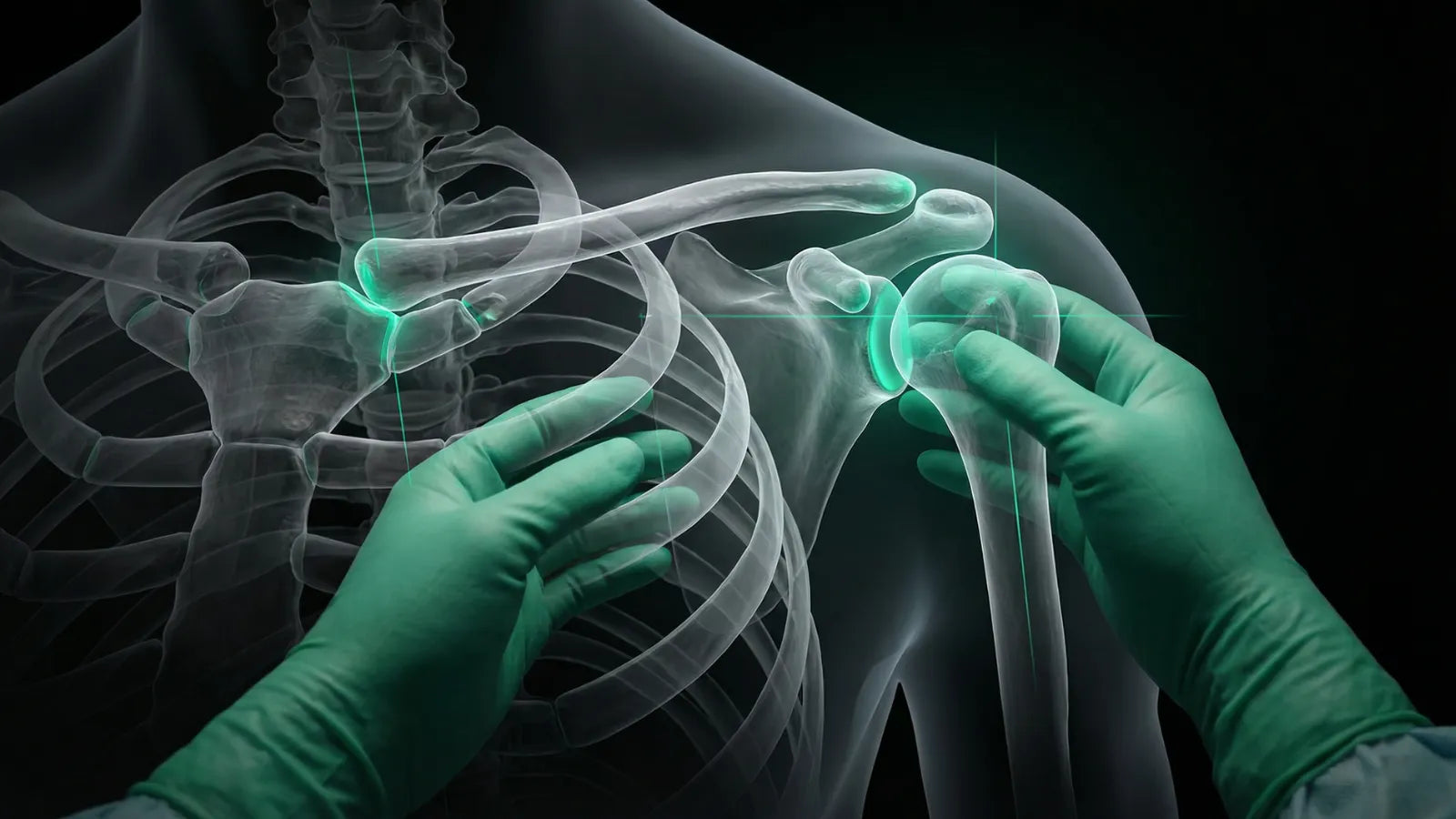
Artificial Intelligence and 3D Planning in Complex Orthopedic Trauma
Orthopedic trauma surgery is undergoing a technological transformation. Artificial intelligence (AI) and 3D printing are no longer distant promises but are increasingly integrated into the pre-operative planning of complex cases. In technically demanding fractures—such as those of the tibial pilon and acetabulum—these tools offer surgeons an additional layer of analysis and predictability even before the patient arrives in the operating room.
How AI contributes to diagnosis and planning
Deep learning algorithms can identify fracture patterns that might go unnoticed in conventional analyses. Based on computed tomography scans, AI performs automated segmentation of bone structures and assists in characterizing the fracture line, providing the surgeon with structured information for choosing the most appropriate approach and implants.
The practical result is more agile and personalized planning: instead of manually interpreting slice by slice, the surgeon receives a data-driven summary, tailored to each patient's anatomical particularities.
In this context, highlights include:
- automated segmentation
- identification of fracture patterns
- support for implant decision-making
- Making the process faster without sacrificing precision.
- The role of 3D printing in surgical practice
3D printing translates digital data into physical objects, making tangible what imaging exams only suggest. From a three-dimensional model of the fracture, the surgeon can assess the geometry of the injury, simulate the reduction of fragments, and pre-contour plates before the operation—reducing the time spent on adjustments on the table.
Customized surgical guides enhance this precision: by accurately guiding the trajectory of screws and performing osteotomies, they contribute to more reproducible results. Studies in the area indicate a measurable reduction in operative time with the use of this approach.
Applications in complex fractures
The utility of these technologies is particularly evident in fractures that combine high anatomical complexity with high technical demand.
- Tibial pilon: 3D printing allows for a detailed visualization of the articular surface, promoting more precise fragment reduction.
- Acetabulum: the support is particularly valuable in the face of complex anatomies, contributing to safer implant positioning.
- Distal humerus: combined use with structural analysis offers a level of detail compatible with the particularities of a complex anatomy.
Practical application in the Brazilian scenario
In Brazil, 3D printing has already found its place in public orthopedics. The 3D Printing and Rehabilitation Technology Center (Centir), linked to the National Institute of Traumatology and Orthopedics (Into) in Rio de Janeiro, is a concrete example of how this technology can operate within the Unified Health System.
The unit features:
- production of about 200 prostheses per year
- significant reduction in manufacturing time (from approximately ten hours to four)
- In addition to prostheses, it enables the production of biomodels and surgical guides used in pre-operative planning, expanding access to personalized solutions for public health network patients.
Future perspectives
The trend is towards increasing integration between already available technologies and new tools that expand their applicability.
Among the main expected advances:
- virtual and augmented reality in training and intraoperative navigation
- automation in 3D model generation
- development of more accessible personalized implants
Still, concrete challenges limit large-scale adoption: the costs associated with the technology, the need for process standardization, and greater clinical validation remain open questions.
Conclusion
The combination of artificial intelligence and 3D printing represents a qualitative shift in how complex orthopedic trauma is planned and executed. By offering greater anatomical predictability and better preparation for intraoperative challenges, these tools expand the safety margin of procedures—for both the surgeon and the patient.

%20and%203D%20printing%20are%20no%20longer%20distant%20promises%20but%20are%20increasingly%20integrated%20into%20the%20pre-oper...)